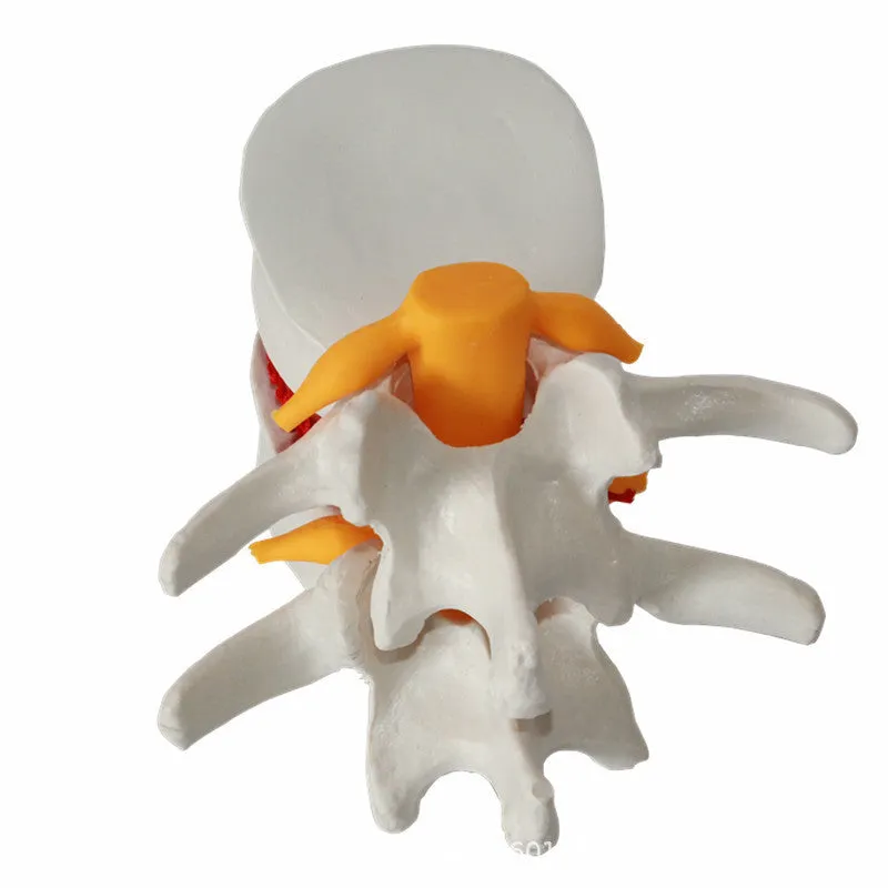
Demonstration model of medical human lumbar intervertebral disc showing herniation pathology, made from durable PVC material measuring 11 x 10 x 7.6 cm with white anatomical colouring for medical education and training purposes.


Demonstration model of medical human lumbar intervertebral disc showing herniation pathology, made from durable PVC material measuring 11 x 10 x 7.6 cm with white anatomical colouring for medical education and training purposes.
Detailed anatomical model showing human lumbar intervertebral disc herniation for medical education

This medical demonstration model provides healthcare educators, students, and practitioners with a tangible representation of lumbar intervertebral disc pathology. Specifically designed to illustrate herniated disc conditions, the three-dimensional format allows for comprehensive examination of spinal anatomy abnormalities. The model serves as an educational bridge between textbook diagrams and actual patient presentations, enabling clearer understanding of disc compression, nerve root impingement, and related anatomical relationships. Medical schools, physiotherapy training programmes, and clinical education departments will find this focused demonstration tool valuable for explaining patient conditions, preparing for surgical procedures, or teaching spinal biomechanics principles.
Features and Construction
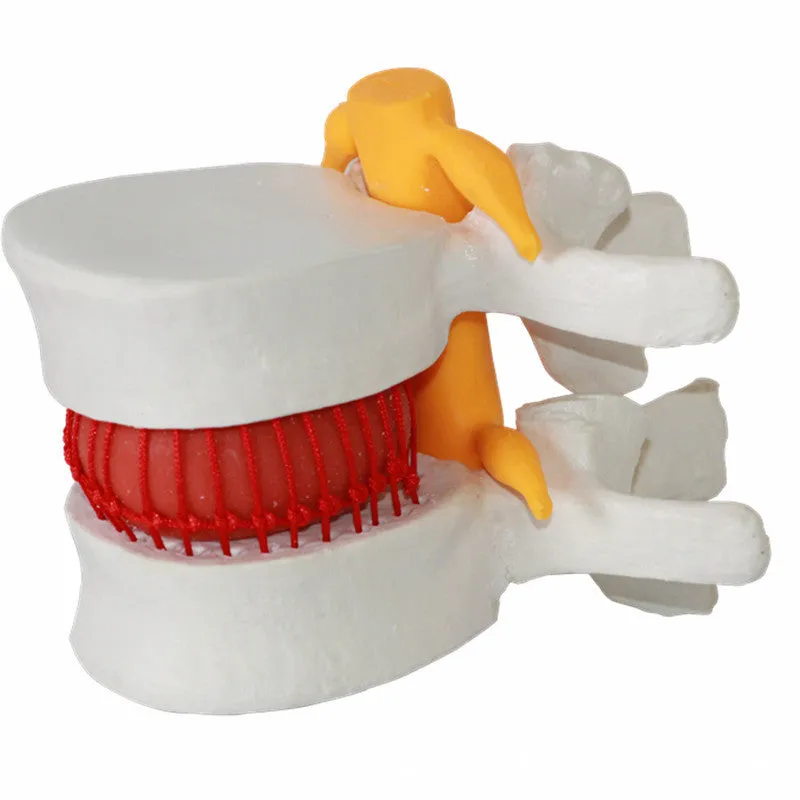
The model's construction prioritises anatomical accuracy and durability for repeated educational use. Each component is designed to show specific aspects of lumbar disc pathology while maintaining structural integrity during handling and demonstration sessions.
Material and Build
Constructed entirely from PVC (Polyvinyl Chloride), this demonstration model offers a rigid yet detailed representation of lumbar intervertebral disc herniation. The white colour scheme provides clear visual differentiation between anatomical structures, allowing educators to point out specific areas of interest during teaching sessions. PVC material ensures the model withstands regular handling in educational environments while maintaining precise anatomical details that illustrate disc pathology. This material choice makes the model suitable for repeated use in classroom settings, clinical demonstrations, and training workshops without significant wear to critical anatomical features.
Size and Practical Fit
Measuring 11 cm in length, 10 cm in width, and 7.6 cm in height, the model provides a substantial yet portable representation of lumbar disc anatomy. These dimensions allow for detailed examination of disc herniation while remaining manageable for single-handed demonstration during lectures or patient consultations. Weighing 0.22 kg, the model offers enough heft to feel substantial during handling without being cumbersome for extended teaching sessions. The compact size facilitates storage in educational cupboards, medical bags, or on clinic shelves when not actively used for demonstration purposes.
Uses and Placement

This focused anatomical model serves multiple educational purposes across healthcare training and clinical environments. Its specific representation of disc pathology makes it particularly valuable for explaining common spinal conditions to various audiences with different learning needs.
Event or Professional Use
In professional medical education settings, this model supports teaching spinal anatomy, pathology, and related clinical presentations. Medical schools can incorporate it into orthopaedic or neurological curriculum modules to illustrate common causes of back pain and sciatica. Physiotherapy training programmes benefit from its clear representation of disc herniation when teaching assessment techniques and treatment rationale. Hospital training departments may use it during staff education sessions to explain MRI or CT scan findings to multidisciplinary teams, helping bridge imaging findings with three-dimensional anatomical understanding.
Everyday Home Use
While primarily designed for professional education, healthcare practitioners may find this model useful for patient education in clinical settings. Chiropractors, osteopaths, and physiotherapists can use it to explain disc conditions to patients, helping them visualise their specific pathology and understand treatment recommendations. The clear white colouring and detailed anatomical representation make complex spinal concepts more accessible to patients with varying levels of medical knowledge. This visual aid can support informed consent discussions before procedures or help patients understand rehabilitation protocols following spinal interventions.
Benefits and Buying Value

This demonstration model offers specific advantages for medical education compared to two-dimensional teaching materials. Its focused design on a single pathology provides targeted learning value that complements broader anatomical models in educational collections.
Reuse and Low Maintenance
The PVC construction requires minimal maintenance while supporting repeated educational use across multiple teaching sessions. Unlike perishable biological specimens or fragile plaster models, this durable representation withstands regular handling by students and educators. Simple cleaning with a damp cloth maintains its appearance without requiring specialised preservation techniques. The model remains stable in various classroom environments without temperature or humidity controls needed for some biological teaching aids. This durability ensures long-term value as an educational investment that serves multiple student cohorts over extended periods.
Why Choose This Product
This specific demonstration model focuses exclusively on lumbar intervertebral disc herniation, providing detailed anatomical representation that general spinal models may not emphasise. The 11 x 10 x 7.6 cm dimensions offer sufficient detail for close examination while remaining portable for varied teaching locations. Unlike generic anatomical models, this specialised representation helps educators explain precise pathological changes associated with disc compression and nerve root involvement. The included packaging provides protective storage that maintains model integrity between educational sessions, supporting organised teaching resource management in busy educational or clinical environments.